In Office And Hospital Services
Ultrasound
Ultrasound imaging is a noninvasive medical test that helps physicians diagnose and treat medical conditions.
Ultrasound imaging uses sound waves to produce pictures of the inside of the body. It is used to help diagnose the causes of pain, swelling and infection in the body's internal organs and to examine a baby in pregnant women and the brain and hips in infants. It's also used to help guide biopsies, diagnose heart conditions, and assess damage after a heart attack. Ultrasound is safe, noninvasive, and does not use ionizing radiation.
This procedure requires little to no special preparation. Your doctor will instruct you on how to prepare, including whether you should refrain from eating or drinking beforehand. Leave jewelry at home and wear loose, comfortable clothing. You may be asked to wear a gown. Ultrasound is safe and painless, and produces pictures of the inside of the body using sound waves. Ultrasound imaging, also called ultrasound scanning or sonography, involves the use of a small transducer (probe) and ultrasound gel placed directly on the skin. High-frequency sound waves are transmitted from the probe through the gel into the body. The transducer collects the sounds that bounce back and a computer then uses those sound waves to create an image. Ultrasound examinations do not use ionizing radiation (as used in x-rays), thus there is no radiation exposure to the patient. Because ultrasound images are captured in real-time, they can show the structure and movement of the body's internal organs, as well as blood flowing through blood vessels.
More advanced ultrasound techniques may be used when a more detailed image is required. These may give the doctor the information necessary to make a diagnosis.
Transvaginal Ultrasound
A transvaginal ultrasound may be done to produce a clearer image. This ultrasound is more likely to be used during the early stages of pregnancy, when capturing a clear image may be more difficult. For this test, a small ultrasound probe is inserted into the vagina. The probe rests against the back of your vagina while the images are captured.
3-D Ultrasound
Unlike a traditional 2-D ultrasound, a 3-D ultrasound allows your doctor to see the width, height, and depth of the fetus and your organs. This ultrasound can be especially helpful in diagnosing any suspected problems during your pregnancy.
A 3-D ultrasound follows the same procedure as a standard ultrasound, but it uses a special probe and software to create the 3-D image. It also requires special training for the technician, so it may not be as widely available.
4-D Ultrasound
A 4-D ultrasound may also be called a dynamic 3-D ultrasound. Unlike other ultrasounds, a 4-D ultrasound creates a moving video of the fetus. It creates a better image of the baby’s face and movements. It also captures highlights and shadows better. This ultrasound is performed similarly to other ultrasounds, but with special equipment.
This procedure requires little to no special preparation. Your doctor will instruct you on how to prepare, including whether you should refrain from eating or drinking beforehand. Leave jewelry at home and wear loose, comfortable clothing. You may be asked to wear a gown. Ultrasound is safe and painless, and produces pictures of the inside of the body using sound waves. Ultrasound imaging, also called ultrasound scanning or sonography, involves the use of a small transducer (probe) and ultrasound gel placed directly on the skin. High-frequency sound waves are transmitted from the probe through the gel into the body. The transducer collects the sounds that bounce back and a computer then uses those sound waves to create an image. Ultrasound examinations do not use ionizing radiation (as used in x-rays), thus there is no radiation exposure to the patient. Because ultrasound images are captured in real-time, they can show the structure and movement of the body's internal organs, as well as blood flowing through blood vessels.
A transvaginal ultrasound may be done to produce a clearer image. This ultrasound is more likely to be used during the early stages of pregnancy, when capturing a clear image may be more difficult. For this test, a small ultrasound probe is inserted into the vagina. The probe rests against the back of your vagina while the images are captured.
Unlike a traditional 2-D ultrasound, a 3-D ultrasound allows your doctor to see the width, height, and depth of the fetus and your organs. This ultrasound can be especially helpful in diagnosing any suspected problems during your pregnancy.
A 3-D ultrasound follows the same procedure as a standard ultrasound, but it uses a special probe and software to create the 3-D image. It also requires special training for the technician, so it may not be as widely available.
A 4-D ultrasound may also be called a dynamic 3-D ultrasound. Unlike other ultrasounds, a 4-D ultrasound creates a moving video of the fetus. It creates a better image of the baby’s face and movements. It also captures highlights and shadows better. This ultrasound is performed similarly to other ultrasounds, but with special equipment.
OB-GYN Services
The term “OB-GYN” refers to the practice of both obstetrics and gynecology or to the doctor who practices both fields of medicine. Some doctors choose to practice only one of these fields. For example, gynecologists only practice gynecology, which focuses on women’s reproductive health.
At Dr. Sherri S. Levin & Associates, we provide comprehensive Obstetrics and Gynecology services. You'll find that within our office we can take care of many of your needs including minimally invasive office surgeries, ultrasound, and a myriad of testing available. Come in and see what we have to offer.
We do not share 'After Hour Calls' with other groups so when you need us after hours you can be certain you will hear from one of the 4 of us and not a physician outside of our group.
Below is a list of some of the OB-GYN services we offer:
Annual exams for all ages
Evaluation of abnormal bleeding and fibroids
Menopausal hormone evaluation and treatment
Urinary incontinence workup and treatment
Pre-pregnancy planning and prenatal care
Infertility workup and treatment
PMS - Premenstrual Syndrome
Acne & abnormal hair growth
Evaluation of birth control methods
Robotic Laparoscopic Hysterectomy
Laparoscopic Bladder Repair & Sling
Laparoscopic removal of the ovaries
Laparoscopic treatment of endometriosis
Treatment of pelvic prolapse
Vaginal reconstruction
Treatment of Dysplasia
Bladder Lift for stress urinary incontinence without mesh
Mesh-less Bladder Lift
Mesh-less Bladder Suspension
Stress Urinary Incontinence
Pelvic Prolapse
Uterine Prolapse
Cervical Prolapse
Cystocele
Rectocele
Vaginal Suspension
Lab Work
Most patients will get a pap smear annually however, there might be reasons to order additional lab work. If you are experiencing some of the following symptoms:
- Fatigue or abnormal weight gain.
- Hair loss or abnormal hair growth
- Acne
Why do some blood tests require fasting?
Everything you eat and drink contains vitamins, proteins, and other nutrients that can cause the related levels in your blood to temporarily spike or drop.
- Fatigue or abnormal weight gain.
- Hair loss or abnormal hair growth
- Acne
Everything you eat and drink contains vitamins, proteins, and other nutrients that can cause the related levels in your blood to temporarily spike or drop.
Endometrial Ablation
Endometrial ablation is a permanent procedure. This procedure is helpful for many women, but isn’t recommended for everyone. Talk to your doctor about whether this is the best option for you.
Your doctor may recommend this procedure if your menstrual periods are extremely heavy and can’t be controlled with medication. Doctors consider menstrual flow to be too heavy if your tampon or sanitary pad is routinely soaked through within two hours.
They may also recommend this procedure if you experience:
- heavy menstrual bleeding that lasts for eight days or longer
- bleeding between periods
- anemia as a result of your period
Endometrial Ablation: What to Expect
- Preparation
- Fertility
- Procedure
- Post-procedure care
- Risks and complications
- Outlook
Who gets endometrial ablation?
Endometrial ablation is a procedure designed to destroy the uterine lining (endometrium).
Your doctor may recommend this procedure if your menstrual periods are extremely heavy and can’t be controlled with medication. Doctors consider menstrual flow to be too heavy if your tampon or sanitary pad is routinely soaked through within two hours.
They may also recommend this procedure if you experience:
- Heavy menstrual bleeding that lasts for eight days or longer
- Bleeding between periods
- Anemia as a result of your period
How to prepare
Prior to scheduling, you’ll discuss your medication history and any allergies you have with your doctor.
If you and your doctor decide to move forward with the procedure, they’ll discuss all aspects of the procedure with you ahead of time. This includes what you should and shouldn’t do in the days and weeks leading up to it.
Standard pre-procedure protocols include:
- Taking a pregnancy test
- Having your IUD removed
- Being tested for endometrial cancer
You may need to have your uterine lining thinned beforehand in order to make the procedure more effective. This may be done with medication or with a dilation and curettage (D and C) procedure.
All endometrial ablation procedures require anesthesia. If general anesthesia is needed, you’ll be instructed to stop eating and drinking eight hours before the procedure.
Additional pre-surgery tests may also be done.
What to expect after the procedure
The type of procedure you have will determine, in part, your post-procedure care and length of recuperation. If you need general anesthesia, your doctor will have you remain in the hospital for several hours after surgery.
No matter what type of procedure you have, you’ll need someone to take you home afterward. You should also bring a sanitary pad with you to wear after the procedure is completed. Talk to your doctor about over-the-counter medication to take for cramps or nausea, and which ones to avoid.
After the procedure, you may experience:
- Increased urination for about a day
- Menstrual-type cramping for several days
- Watery, bloody vaginal discharge for several weeks
- Nausea
You should seek emergency medical attention if you experience:
- Foul-smelling discharge
- Fever
- Chills
- Trouble urinating
- Heavy bleeding
- Extreme abdominal cramping
- Preparation
- Fertility
- Procedure
- Post-procedure care
- Risks and complications
- Outlook
Endometrial ablation is a procedure designed to destroy the uterine lining (endometrium).
Your doctor may recommend this procedure if your menstrual periods are extremely heavy and can’t be controlled with medication. Doctors consider menstrual flow to be too heavy if your tampon or sanitary pad is routinely soaked through within two hours.
They may also recommend this procedure if you experience:
- Heavy menstrual bleeding that lasts for eight days or longer
- Bleeding between periods
- Anemia as a result of your period
Prior to scheduling, you’ll discuss your medication history and any allergies you have with your doctor.
If you and your doctor decide to move forward with the procedure, they’ll discuss all aspects of the procedure with you ahead of time. This includes what you should and shouldn’t do in the days and weeks leading up to it.
Standard pre-procedure protocols include:
- Taking a pregnancy test
- Having your IUD removed
- Being tested for endometrial cancer
The type of procedure you have will determine, in part, your post-procedure care and length of recuperation. If you need general anesthesia, your doctor will have you remain in the hospital for several hours after surgery.
No matter what type of procedure you have, you’ll need someone to take you home afterward. You should also bring a sanitary pad with you to wear after the procedure is completed. Talk to your doctor about over-the-counter medication to take for cramps or nausea, and which ones to avoid.
- Increased urination for about a day
- Menstrual-type cramping for several days
- Watery, bloody vaginal discharge for several weeks
- Nausea
- Foul-smelling discharge
- Fever
- Chills
- Trouble urinating
- Heavy bleeding
- Extreme abdominal cramping
Bone Density Testing
Bone Density, also known as Bone Mineral Density (BMD)
If you have osteopenia, you have lower bone density than normal. Your bone density peaks when you’re about 35 years old.
Bone mineral density (BMD) is the measurement of how much bone mineral is in your bones. Your BMD estimates the chances of breaking a bone from a normal activity. People who have osteopenia have a lower BMD than normal, but it’s not a disease.
However, having osteopenia does increase your chances of developing osteoporosis. This bone disease causes fractures, stooped posture, and can lead to severe pain.
You can take action to prevent osteopenia. The right exercise and food choices may help keep your bones strong. If you have osteopenia, ask your doctor about how to keep it from worsening so you can prevent osteoporosis
Aging is the most common risk factor for osteopenia. After your bone mass peaks, your body breaks down old bone faster than it builds new bone. That means you lose some bone density. Women lose bone more quickly after menopause. If you lose too much, your bone mass may drop low enough to be considered osteopenia.
About half of Americans older than age 50 get osteopenia. The more of these risk factors you have, the higher your risk is:
- Being female, with small-boned women of Asian and Caucasian descent having the highest risk
- Family history of low BMD
- Being older than age 50
- Menopause before age 45
- Removal of ovaries before menopause
- Not getting enough exercise
- A poor diet, especially one lacking calcium and vitamin D
- Smoking or using other forms of tobacco
- Drinking too much alcohol or caffeine
- Taking prednisone or phenytoin
The goal of treatment is to keep osteopenia from progressing into osteoporosis.
The first part of treatment involves diet and exercise choices. The risk of breaking a bone when you have osteopenia is fairly small, so doctors don’t usually prescribe medicine unless your BMD is very close to the osteoporosis level. They may talk to you about taking a calcium or vitamin D supplement, although generally it’s better to get enough of each from your diet.
Bone mineral density (BMD) is the measurement of how much bone mineral is in your bones. Your BMD estimates the chances of breaking a bone from a normal activity. People who have osteopenia have a lower BMD than normal, but it’s not a disease.
However, having osteopenia does increase your chances of developing osteoporosis. This bone disease causes fractures, stooped posture, and can lead to severe pain.
You can take action to prevent osteopenia. The right exercise and food choices may help keep your bones strong. If you have osteopenia, ask your doctor about how to keep it from worsening so you can prevent osteoporosis
About half of Americans older than age 50 get osteopenia. The more of these risk factors you have, the higher your risk is:
- Being female, with small-boned women of Asian and Caucasian descent having the highest risk
- Family history of low BMD
- Being older than age 50
- Menopause before age 45
- Removal of ovaries before menopause
- Not getting enough exercise
- A poor diet, especially one lacking calcium and vitamin D
- Smoking or using other forms of tobacco
- Drinking too much alcohol or caffeine
- Taking prednisone or phenytoin
The goal of treatment is to keep osteopenia from progressing into osteoporosis.
The first part of treatment involves diet and exercise choices. The risk of breaking a bone when you have osteopenia is fairly small, so doctors don’t usually prescribe medicine unless your BMD is very close to the osteoporosis level. They may talk to you about taking a calcium or vitamin D supplement, although generally it’s better to get enough of each from your diet.
The first part of treatment involves diet and exercise choices. The risk of breaking a bone when you have osteopenia is fairly small, so doctors don’t usually prescribe medicine unless your BMD is very close to the osteoporosis level. They may talk to you about taking a calcium or vitamin D supplement, although generally it’s better to get enough of each from your diet.
Laparoscopic Surgery
Laparoscopy, also known as diagnostic laparoscopy, is a surgical diagnostic procedure used to examine the organs inside the abdomen. It’s a low-risk, minimally invasive procedure that requires only small incisions.
Laparoscopy uses an instrument called a laparoscope to look at the abdominal organs. A laparoscope is a long, thin tube with a high-intensity light and a high-resolution camera at the front. The instrument is inserted through an incision in the abdominal wall. As it moves along, the camera sends images to a video monitor.
Laparoscopy allows your doctor to see inside your body in real time, without open surgery. Your doctor also can obtain biopsy samples during this procedure.
Laparoscopy is often used to identify and diagnose the source of pelvic or abdominal pain. It’s usually performed when noninvasive methods are unable to help with diagnosis.
In many cases, abdominal problems can also be diagnosed with imaging techniques such as:
- Ultrasound, which uses high-frequency sound waves to create images of the body
- CT scan, which is a series of special X-rays that take cross-sectional images of the body
- MRI scan, which uses magnets and radio waves to produce images of the body
Laparoscopy is performed when these tests don’t provide enough information or insight for a diagnosis. The procedure may also be used to take a biopsy, or sample of tissue, from a particular organ in the abdomen.
Laparoscopy is usually done as an outpatient procedure where you are always be given given general anesthesia. This means that you’ll be able to go home the same day as your surgery. It may be performed in a hospital or an outpatient surgical center.
You’ll likely be given general anesthesia for this type of surgery. This means that you’ll sleep through the procedure and won’t feel any pain. To achieve general anesthesia, an intravenous (IV) line is inserted in one of your veins. Through the IV, your anesthesiologist can give you special medications and well as provide hydration with fluids.
During laparoscopy, the surgeon makes an incision at your belly button, and then inserts a small tube called a cannula. The cannula is used to inflate your abdomen with carbon dioxide gas. This gas allows your doctor to see your abdominal organs more clearly.
Once your abdomen is inflated, the surgeon inserts the laparoscope through the incision. The camera attached to the laparoscope displays the images on a screen, allowing your organs to be viewed in real time.
The number and size of incisions depends upon what specific medical conditions your surgeon is attempting to confirm or rule out. Generally, you get from 1 to 5 incisions that are each between 1 and 2 centimeters in length. These incisions allow other instruments to be inserted. For example, your surgeon may need to use another surgical tool to perform a biopsy. During a biopsy, they take a small sample of tissue from an organ to be evaluated.
After the procedure is done, the instruments are removed. Your incisions are then closed with stitches or surgical tape. Bandages may be placed over the incisions.
Laparoscopy uses an instrument called a laparoscope to look at the abdominal organs. A laparoscope is a long, thin tube with a high-intensity light and a high-resolution camera at the front. The instrument is inserted through an incision in the abdominal wall. As it moves along, the camera sends images to a video monitor.
Laparoscopy allows your doctor to see inside your body in real time, without open surgery. Your doctor also can obtain biopsy samples during this procedure.
In many cases, abdominal problems can also be diagnosed with imaging techniques such as:
- Ultrasound, which uses high-frequency sound waves to create images of the body
- CT scan, which is a series of special X-rays that take cross-sectional images of the body
- MRI scan, which uses magnets and radio waves to produce images of the body
You’ll likely be given general anesthesia for this type of surgery. This means that you’ll sleep through the procedure and won’t feel any pain. To achieve general anesthesia, an intravenous (IV) line is inserted in one of your veins. Through the IV, your anesthesiologist can give you special medications and well as provide hydration with fluids.
During laparoscopy, the surgeon makes an incision at your belly button, and then inserts a small tube called a cannula. The cannula is used to inflate your abdomen with carbon dioxide gas. This gas allows your doctor to see your abdominal organs more clearly.
Once your abdomen is inflated, the surgeon inserts the laparoscope through the incision. The camera attached to the laparoscope displays the images on a screen, allowing your organs to be viewed in real time.
The number and size of incisions depends upon what specific medical conditions your surgeon is attempting to confirm or rule out. Generally, you get from 1 to 5 incisions that are each between 1 and 2 centimeters in length. These incisions allow other instruments to be inserted. For example, your surgeon may need to use another surgical tool to perform a biopsy. During a biopsy, they take a small sample of tissue from an organ to be evaluated.
After the procedure is done, the instruments are removed. Your incisions are then closed with stitches or surgical tape. Bandages may be placed over the incisions.
Another Day In ParadiseSpecial Rejuvenating Treatments To Beautify You.
da Vinci Robotics
Got a Question? We’re here to help.
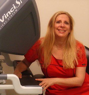
The da Vinci surgical system is a breakthrough surgical technology that allows for a minimally invasive approach to common gynecologic surgical conditions. Dr. Sherri Levin has been performing da Vinci assisted surgeries since 2007 and has completed over 1,600 cases as of 2019. She previously taught doctors from around the country the appropriate utilization of the da Vinci system in routine and advanced cases. As a result of her experience and expertise, she was asked to appear on The Doctors as an expert on this surgical approach in 2008. Most recently, she was designated a da Vinci Gynecologic Epicenter. Dr. Anne Gonzalez and Dr. Amelie Chu are also certified daVinci robotic surgeons.
We believe that almost anyone is a candidate for a da Vinci surgery and continue to be amazed by the phenomenal results that this new surgical system provides. Listed below are some common questions and answers on the da Vinci system...
We believe that almost anyone is a candidate for a da Vinci surgery and continue to be amazed by the phenomenal results that this new surgical system provides. Listed below are some common questions and answers on the da Vinci system...
Dr. Sherri Levin has been performing da Vinci assisted surgeries since 2007 and has completed over 1,600 da Vinci cases as of 2019.
Robotic Patient Testimonials
Dr. Sherri Levin has been performing da Vinci assisted surgeries
since 2007 and has completed over 1,600 cases as of 2019.
“The surgery was a great relief to me. I am not in pain anymore. I used to dread my monthly cycle because of the heavy bleeding. My recovery time was quick, and I am feeling better than ever.”
Tracy W
Robotic Assisted
Surgery Patient
“Recovery time was only about 3 days, and then able to do more every day. Important to have help for about 1.5 to 2 weeks. I was briefed and received information so I knew more or less, what to expect. Hospital and staff were great and I was out showing homes 4 days after my surgery...with my husband driving.”
Christine R
Robotic Assisted
Surgery Patient
“I suffered with Heavy periods for 25+ years and High Pain Cramps. My recovery period was minimal, and my pain, compared to my prior pain. I feel like a new, vibrant, high-energy woman. Should have done this several years ago.”
Tony S
Robotic Assisted
Surgery Patient
“Surgery was explained in detail.
I had total confidence in Dr. Levin.
I am so glad I had the surgery,
I am so glad I had the surgery,
I feel like a new person.”
Nancy H
Robotic Assisted
Surgery Patient
“Very pleased with everything and am feeling like a new woman.
Feel 100% better. Thanks Dr. L."
Jeri F
Robotic Assisted
Surgery Patient
“I had the surgery late Wed. afternoon and by the evening I was awake and feeling pretty good. By the next morning I was up and going to the bathroom and eating. I left Thursday AM and continued to improve. Moderate pain for 3 more days, treated with medication. By Sunday I was a new person. I was no longer on pain meds and my pain was low to none. By that Friday I was back to normal. I haven’t felt this good in a year!”
Deana S
Robotic Assisted
Surgery Patient